

A doctor’s simple question
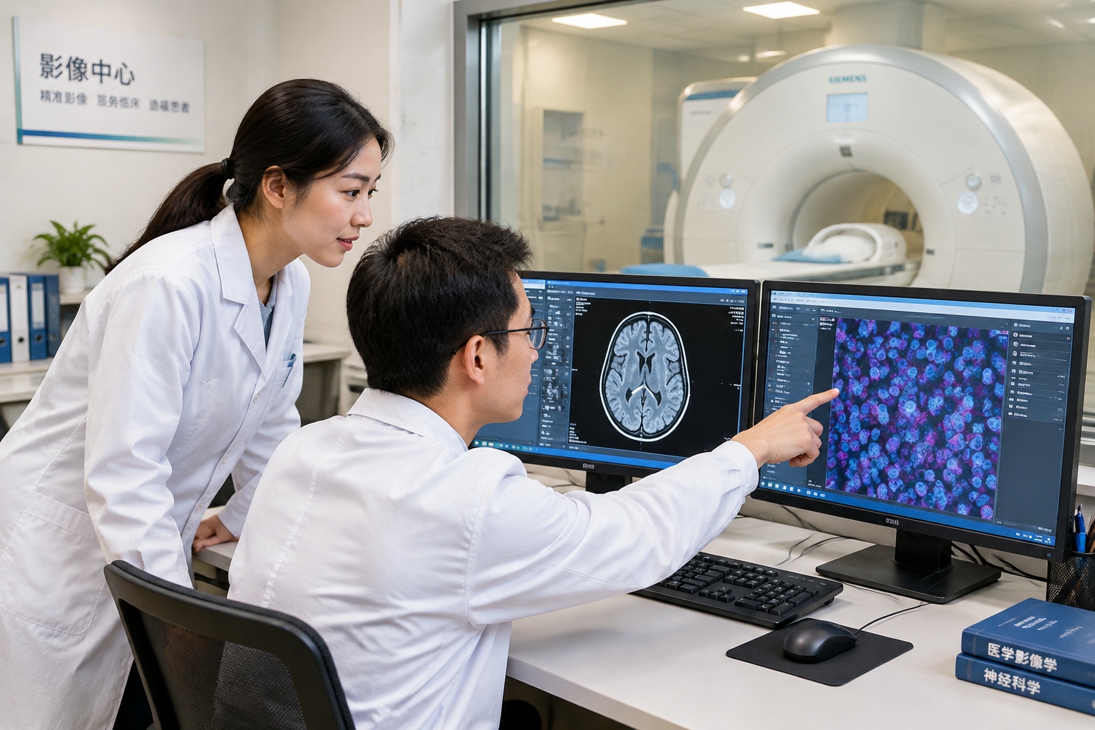
A radiologist points at a gray smudge on a CT scan. The patient asks the obvious question: is it cancer? The scan can show size, shape and location. It cannot easily show what the cells inside that smudge are doing.
That gap—between seeing a shadow and understanding its biology—is the everyday problem behind a large, slightly intimidating piece of Chinese science infrastructure: the Multimodal Cross-scale Biomedical Imaging Facility in Beijing’s Huairou Science City.
The name sounds like a government document. The idea is easier. Doctors and scientists want to follow a disease from molecules to cells to tissue to organs, without cutting the patient open for every answer. No single scanner can do that. So the facility connects many kinds of imaging, and the computing needed to make their pictures talk to each other.
Why one camera is never enough
Think about the camera on your phone. A wide shot shows the whole street. A macro shot shows a fly on a windowsill. You cannot get both in one frame at the same level of detail. Biomedical imaging has the same trade-off: resolution versus field of view.
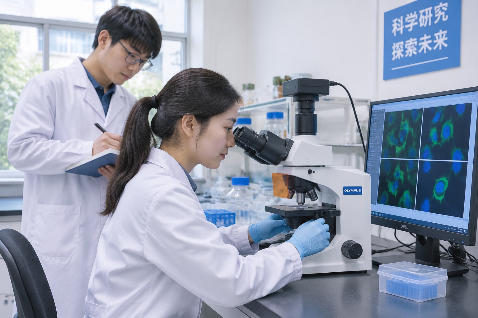
A whole-body MRI can show a liver, a heart or a brain tumor in context. It sees centimeters, sometimes millimeters. A super-resolution fluorescence microscope can see structures inside a single cell, down to tens of nanometers. A cryo-electron microscope can reveal the shape of proteins frozen in place, at near-atomic scale. Each machine answers a different question.
The contrasts differ too. CT uses X-rays and shows dense tissue well. MRI uses magnetic fields and radio waves, and is good for soft tissue. PET uses a radioactive tracer to show metabolism. Ultrasound uses sound waves and is cheap, fast and everywhere. Fluorescence microscopy uses light and dyes to label specific molecules. None of these is a universal truth machine. Each is a partial view.
“Multimodal” means using several of these methods on the same biological problem. “Cross-scale” means linking what they see across size ranges—molecule, cell, tissue, organ, whole body. In practice, that is less like one giant machine and more like a well-run airport: different runways, different aircraft, one control tower.
What the facility actually is
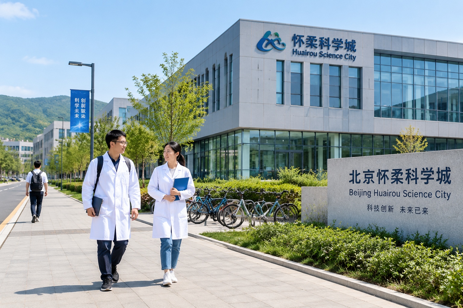
Chinese official materials describe the facility as a national major science and technology infrastructure led by Peking University, located in Huairou Science City, a research cluster north of Beijing. Public descriptions list a range of instruments and methods, including cryo-electron microscopy, super-resolution fluorescence imaging, medical imaging such as MRI and PET, and data-processing platforms. The exact equipment list is less important for a general reader than the design goal: make separate views of biology comparable.
That last point is the hard part. You cannot put a living person inside an electron microscope. You cannot squeeze a whole-body PET scanner into a cell biology lab. So the facility works with samples, model organisms and human scans in a coordinated way. A team might stain a tumor sample with fluorescent markers, image it under a microscope, then use electron microscopy to zoom into the same region. Another project might compare an animal model with human MRI and PET data. The images are registered, scaled and stored so that a researcher can move between them.
This is also why the facility needs more than scanners. Sample preparation, quality control, data storage, computing and expert staff matter as much as the hardware. A beautiful image that cannot be reproduced or compared is not much use.
There is another practical detail: biological samples change. A tissue slice dries, a cell moves, a mouse gets older. Cross-scale work often means finding the same small region twice, on different machines, under different conditions. That sounds boring. It is also where many projects succeed or fail.
Where it could matter: cancer, heart and brain, drugs
Cancer is the most obvious case. A standard scan can show a lump. It may not show whether the lump is aggressive, whether it has certain receptors, or how it is interacting with immune cells. Multimodal imaging can combine anatomy with metabolism, molecular markers and tissue structure. For early detection, the hope is not just to find smaller spots, but to tell which ones need treatment and which can be watched.
Heart and brain disease offer a similar promise. In stroke, doctors need to know quickly whether a blocked vessel can be reopened, and which brain tissue is still salvageable. In brain science, researchers want to link molecular changes with circuits and behavior. In drug development, imaging can help show whether a drug reaches its target and whether it changes the biology as intended—before a large and expensive clinical trial. That could reduce the cost of failure, or at least make failure happen earlier.
None of this is automatic. A powerful image is not a diagnosis. It becomes useful only after clinical studies show that doctors make better decisions with it, and after regulators, hospitals and payment systems accept it.
Who gets to use it?
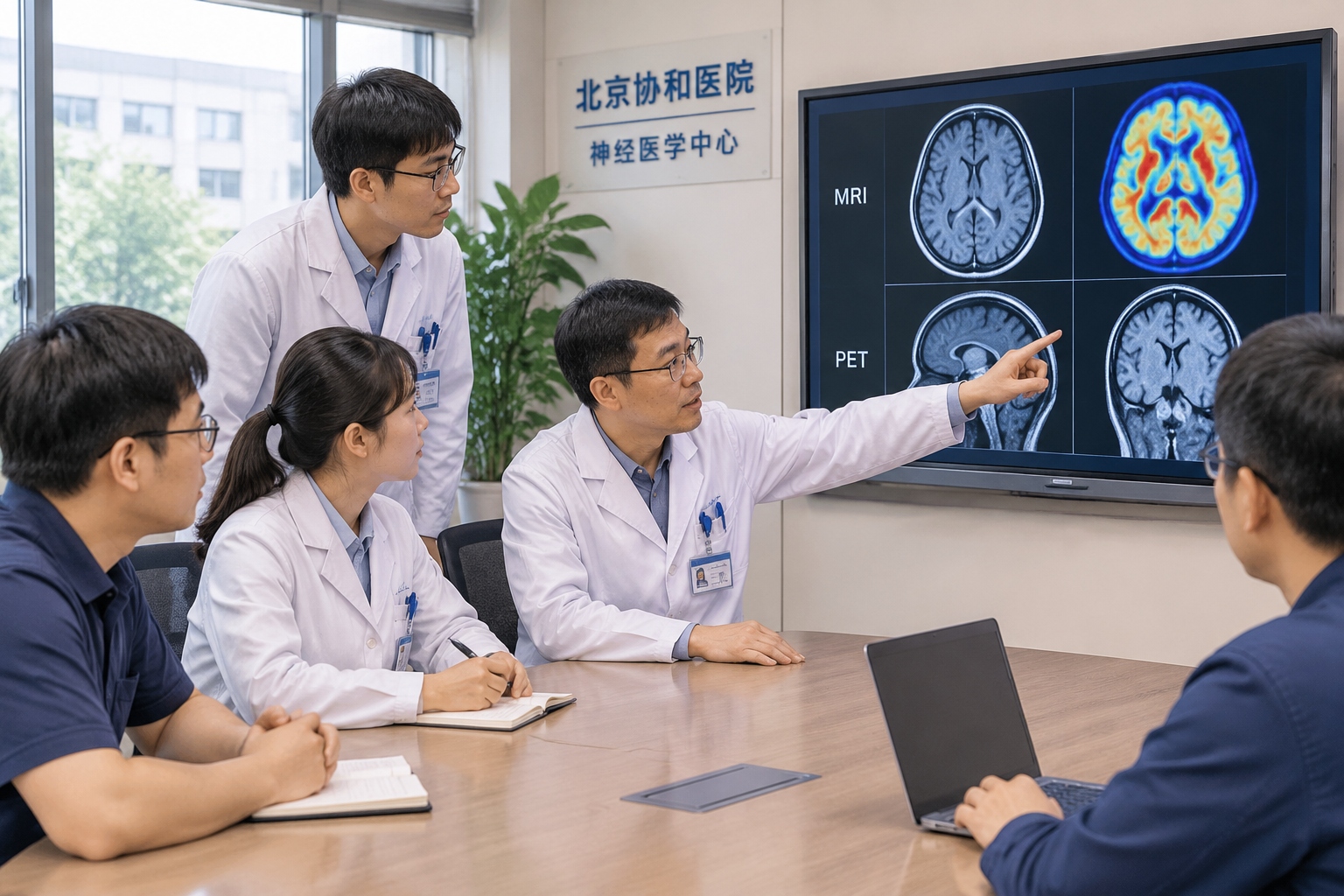
A facility like this is too expensive to be a private toy. The usual model is shared access. Research teams submit proposals. A scientific review panel judges the idea and the feasibility. Approved projects get instrument time, technical support and help with data. Users may include university labs, hospitals and companies. Many major facilities charge user fees, often with subsidies for public research, and require results to be published or otherwise shared.
For overseas readers, the closest analogy is a synchrotron or a national microscopy center: not a walk-in clinic, but a scheduled research service. A patient does not book an appointment at Huairou. A doctor might join a research project, or a hospital might send samples and scans through a collaboration.
Open sharing is a real test for any big science project. It is easy to list instruments on a website. It is harder to make the application process fair, the schedules reliable and the data usable for teams outside the founding institutions.
The honest limits
Chinese official descriptions sometimes use phrases like “international advanced” or “world-leading.” Those claims should be read carefully. The right measure is not a press release but peer-reviewed papers, reproduced methods, clinical trials and practical uptime. A facility can be excellent at some scales and still developing at others. It can have impressive instruments and a long queue for access.
There are also ordinary constraints: cost, trained staff, data privacy, and the gap between a research scan and a routine hospital scan. A technique that takes hours in a quiet lab may not fit a busy emergency department. A contrast agent that works in mice may fail in people. The path from a striking image to a change in medical practice is slow, and that is normal.
What it means for an ordinary person
Your next hospital visit probably will not involve a tour of a cross-scale imaging facility. The benefit arrives indirectly. It might be a better marker for an aggressive tumor, a scan protocol that shortens a procedure, or a drug trial that fails earlier and cheaper. It might be a brain study that changes how doctors think about a disease.
The most useful way to judge this kind of infrastructure is not to ask whether it sounds futuristic. Ask a simpler question: can a doctor somewhere make a decision with more confidence, and with less invasion, because of what this facility helped show? If the answer becomes yes, the long name on the building will have earned its place.